
InVivoPlus anti-mouse PD-1 (CD279)
Product Description
Specifications
| Isotype | Armenian hamster IgG |
|---|---|
| Recommended Isotype Control(s) | InVivoPlus polyclonal Armenian hamster IgG |
| Recommended Dilution Buffer | InVivoPure pH 6.5 Dilution Buffer |
| Conjugation | This product is unconjugated. Conjugation is available via our Antibody Conjugation Services. |
| Immunogen | Syrian Hamster BKH cells transfected with mouse PD-1 cDNA |
| Reported Applications |
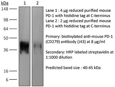
in vivo blocking of PD-1/PD-L signaling in vitro PD-1 neutralization Western blot |
| Formulation |
PBS, pH 6.5 Contains no stabilizers or preservatives |
| Endotoxin* |
≤0.5EU/mg (≤0.0005EU/μg) Determined by LAL assay |
| Aggregation* |
<5% Determined by SEC |
| Purity |
≥95% Determined by SDS-PAGE |
| Sterility | 0.2 µm filtration |
| Production | Purified from cell culture supernatant in an animal-free facility |
| Purification | Protein G |
| RRID | AB_1107747 |
| Molecular Weight | 150 kDa |
| Murine Pathogen Tests* |
Ectromelia/Mousepox Virus: Negative Hantavirus: Negative K Virus: Negative Lactate Dehydrogenase-Elevating Virus: Negative Lymphocytic Choriomeningitis virus: Negative Mouse Adenovirus: Negative Mouse Cytomegalovirus: Negative Mouse Hepatitis Virus: Negative Mouse Minute Virus: Negative Mouse Norovirus: Negative Mouse Parvovirus: Negative Mouse Rotavirus: Negative Mycoplasma Pulmonis: Negative Pneumonia Virus of Mice: Negative Polyoma Virus: Negative Reovirus Screen: Negative Sendai Virus: Negative Theiler’s Murine Encephalomyelitis: Negative |
| Storage | The antibody solution should be stored at the stock concentration at 4°C. Do not freeze. |
| Need a Custom Formulation? | See All Antibody Customization Options |
Application References
-
Park, S. J., et al (2014). "Negative role of inducible PD-1 on survival of activated dendritic cells" J Leukoc Biol 95(4): 621-629.
PubMed
PD-1 is a well-established negative regulator of T cell responses by inhibiting proliferation and cytokine production of T cells via interaction with its ligands, B7-H1 (PD-L1) and B7-DC (PD-L2), expressed on non-T cells. Recently, PD-1 was found to be expressed in innate cells, including activated DCs, and plays roles in suppressing production of inflammatory cytokines. In this study, we demonstrate that PD-1 KO DCs exhibited prolonged longevity compared with WT DCs in the dLNs after transfer of DCs into hind footpads. Interestingly, upon LPS stimulation, WT DCs increased the expression of PD-1 and started to undergo apoptosis. DCs, in spleen of LPS-injected PD-1 KO mice, were more resistant to LPS-mediated apoptosis in vivo than WT controls. Moreover, treatment of blocking anti-PD-1 mAb during DC maturation resulted in enhanced DC survival, suggesting that PD-1:PD-L interactions are involved in DC apoptosis. As a result, PD-1-deficient DCs augmented T cell responses in terms of antigen-specific IFN-gamma production and proliferation of CD4 and CD8 T cells to a greater degree than WT DCs. Moreover, PD-1 KO DCs exhibited increased MAPK1 and CD40-CD40L signaling, suggesting a possible mechanism for enhanced DC survival in the absence of PD-1 expression. Taken together, our findings further extend the function of PD-1, which plays an important role in apoptosis of activated DCs and provides important implications for PD-1-mediated immune regulation.
-
Rabenstein, H., et al (2014). "Differential kinetics of antigen dependency of CD4+ and CD8+ T cells" J Immunol 192(8): 3507-3517.
PubMed
Ag recognition via the TCR is necessary for the expansion of specific T cells that then contribute to adaptive immunity as effector and memory cells. Because CD4+ and CD8+ T cells differ in terms of their priming APCs and MHC ligands we compared their requirements of Ag persistence during their expansion phase side by side. Proliferation and effector differentiation of TCR transgenic and polyclonal mouse T cells were thus analyzed after transient and continuous TCR signals. Following equally strong stimulation, CD4+ T cell proliferation depended on prolonged Ag presence, whereas CD8+ T cells were able to divide and differentiate into effector cells despite discontinued Ag presentation. CD4+ T cell proliferation was neither affected by Th lineage or memory differentiation nor blocked by coinhibitory signals or missing inflammatory stimuli. Continued CD8+ T cell proliferation was truly independent of self-peptide/MHC-derived signals. The subset divergence was also illustrated by surprisingly broad transcriptional differences supporting a stronger propensity of CD8+ T cells to programmed expansion. These T cell data indicate an intrinsic difference between CD4+ and CD8+ T cells regarding the processing of TCR signals for proliferation. We also found that the presentation of a MHC class II-restricted peptide is more efficiently prolonged by dendritic cell activation in vivo than a class I bound one. In summary, our data demonstrate that CD4+ T cells require continuous stimulation for clonal expansion, whereas CD8+ T cells can divide following a much shorter TCR signal.
-
Noval Rivas, M., et al (2009). "Reviving function in CD4+ T cells adapted to persistent systemic antigen" J Immunol 183(7): 4284-4291.
PubMed
In bone marrow-transplanted patients, chronic graft-versus-host disease is a complication that results from the persistent stimulation of recipient minor histocompatibility Ag (mHA)-specific T cells contained within the graft. In this study, we developed a mouse model where persistent stimulation of donor T cells by recipient’s mHA led to multiorgan T cell infiltration. Exposure to systemic mHA, however, deeply modified T cell function and chronically stimulated T cells developed a long-lasting state of unresponsiveness, or immune adaptation, characterized by their inability to mediate organ immune damages in vivo. However, analysis of the gene expression profile of adapted CD4+ T cells revealed the specific coexpression of genes known to promote differentiation and function of Th1 effector cells as well as genes coding for proteins that control T cell activity, such as cell surface-negative costimulatory molecules and regulatory cytokines. Strikingly, blockade of negative costimulation abolished T cell adaptation and stimulated strong IFN-gamma production and severe multiorgan wasting disease. Negative costimulation was also shown to control lethal LPS-induced toxic shock in mice with adapted T cells, as well as the capacity of adapted T cells to reject skin graft. Our results demonstrate that negative costimulation is the molecular mechanism used by CD4+ T cells to adapt their activity in response to persistent antigenic stimulation. The effector function of CD4+ T cells that have adapted to chronic Ag presentation can be activated by stimuli strong enough to overcome regulatory signals delivered to the T cells by negative costimulation.
-
Sarraj, B., et al (2014). "Impaired selectin-dependent leukocyte recruitment induces T-cell exhaustion and prevents chronic allograft vasculopathy and rejection" Proc Natl Acad Sci U S A 111(33): 12145-12150.
PubMed
Selectin-selectin ligand interactions mediate the initial steps in leukocyte migration, an integral part of immune responses. Fucosyltransferase-VII (FucT-VII), encoded by Fut7, is essential for biosynthesis of selectin ligands. In an established model of cardiac allograft vasculopathy and chronic rejection, Fut7(-/-) recipients exhibited long-term graft survival with minimal vasculopathy compared with WT controls. Graft survival was associated with CD4 T-cell exhaustion in the periphery, characterized by impaired effector cytokine production, defective proliferation, increased expression of inhibitory receptors programmed death-1 (PD-1) and T cell Ig- and mucin-domain-containing molecule-3 (Tim-3), low levels of IL-7Ralpha on CD4 T cells, and reduced migration of polyfunctional CD4 memory T cells to the allograft. Blocking PD-1 triggered rejection only in Fut7(-/-) recipients, whereas depleting regulatory T cells had no effect in either Fut7(-/-) or WT recipients. Adoptive transfer experiments confirmed that this CD4 T cell-exhausted phenotype is seen primarily in Fut7(-/-) CD4 T cells. These data suggest that impaired leukocyte recruitment is a novel mechanism leading to CD4 T-cell exhaustion. Our experimental system serves as an excellent model to study CD4 T-cell exhaustion as a dominant mechanism of transplant tolerance. Further, targeting FucT-VII may serve as a promising strategy to prevent chronic allograft rejection and promote tolerance.
Product Citations
-
WNT pathway inhibition sensitizes HAT1-high lung cancers to treatment with PD-1 inhibitors.
In Cancer Cell Int on 24 October 2025 by Yang, T., Xie, Z., et al.
PubMed
Immunotherapies change the paradigm of current pulmonary oncological clinics, although majority of patients fail to benefit from these treatment modalities. HAT1 overexpression is frequently diagnosed in lung cancer patients. Effective immunotherapeutic scheme remains to be determined for this portion of patients.
-
Gut-derived Faecalibaculum rodentium exerts anti-cancer effects on colorectal cancer by modulating PDPN-CLEC-2 signaling pathway.
In mSystems on 19 August 2025 by Yang, X., Liu, M., et al.
PubMed
Dynamic changes in microbial composition are related to the progression of colorectal cancer (CRC); however, specific microbes with anti-CRC properties have not been identified. Here, we aimed to determine the effect of Faecalibaculum rodentium on CRC progression. In this study, the effect of F. rodentium was assessed in an azoxymethane (AOM)/dextran sulfate sodium salt (DSS)-induced CRC mouse model as well as in human CRC cell lines. Changes in anti-tumor immune responses were evaluated by flow cytometry. The functional metabolites produced by F. rodentium were identified by gas chromatography‒mass spectrometry (GC‒MS). The subcutaneously injected BALB/c mice with CT26 CRC cells were used to evaluate the effect of acetate produced by F. rodentium. We found that supplementation with F. rodentium suppressed CRC in an AOM/DSS-induced CRC mouse model as well as in the human CRC cell lines SW620 and HT-29. GC‒MS revealed that acetate is a functional tumor-suppressive metabolite produced by F. rodentium, and the tumor volume and tumor weight in the CRC model were inhibited by acetate administration. Mechanistically, F. rodentium produced acetate, which suppressed the expression of PDPN on CD8+ T cells and that of C-type lectin-like receptor 2 (CLEC-2) on tumor cells. As a consequence, CD8+ T-cell immunity was increased both in vivo and in vitro. In addition, we revealed that the acetate produced by F. rodentium can modulate podoplanin (PDPN)/CLEC-2/PI3K/AKT/mTOR signaling in CRC. In summary, F. rodentium produced acetate inhibits CRC by promoting CD8+ T-cell immunity and modulating the PDPN-CLEC-2 pathway. Our data indicate that F. rodentium is a potential probiotic for cancer prevention and treatment.IMPORTANCEThis study identified Faecalibaculum rodentium as a novel probiotic that suppresses colorectal cancer (CRC) via acetate, which enhances CD8+ T-cell immunity by blocking PDPN-CLEC-2 and induces tumor cell apoptosis through PI3K/AKT/mTOR pathway. These findings validated F. rodentium and acetate as dual-action therapeutic candidates through immune response activation and intrinsic tumor targeting by murine models and human CRC cell lines, thereby providing innovative strategies for CRC treatment.
-
Caerin 1.1/1.9-mediated antitumor immunity depends on IFNAR-Stat1 signalling of tumour infiltrating macrophage by autocrine IFNα and is enhanced by CD47 blockade.
In Sci Rep on 30 January 2025 by Li, J., Luo, Y., et al.
PubMed
Previously, we demonstrated that natural host-defence peptide caerin 1.1/caerin 1.9 (F1/F3) increases the efficacy of anti-PD-1 and therapeutic vaccine, in a HPV16 + TC-1 tumour model, but the anti-tumor mechanism of F1/F3 is still unclear. In this study, we explored the impact of F1/F3 on the tumor microenvironment in a transplanted B16 melanoma model, and further investigated the mechanism of action of F1/F3 using monoclonal antibodies to deplete relevant cells, gene knockout mice and flow cytometry. We show that F1/F3 is able to inhibit the growth of melanoma B16 tumour cells both in vitro and in vivo. Depletion of macrophages, blockade of IFNα receptor, and Stat1 inhibition each abolishes F1/F3-mediated antitumor responses. Subsequent analysis reveals that F1/F3 increases the tumour infiltration of inflammatory macrophages, upregulates the level of IFNα receptor, and promotes the secretion of IFNα by macrophages. Interestingly, F1/F3 upregulates CD47 level on tumour cells; and blocking CD47 increases F1/F3-mediated antitumor responses. Furthermore, F1/F3 intratumor injection, CD47 blockade, and therapeutic vaccination significantly increases the survival time of B16 tumour-bearing mice. These results indicate that F1/F3 may be effective to improve the efficacy of ICB and therapeutic vaccine-based immunotherapy for human epithelial cancers and warrants consideration for clinical trials.
-
Suppressing PD-L1 Expression via AURKA Kinase Inhibition Enhances Natural Killer Cell-Mediated Cytotoxicity against Glioblastoma.
In Cells on 6 July 2024 by Nguyen, T. T. T., Gao, Q., et al.
PubMed
Immunotherapies have shown significant promise as an impactful strategy in cancer treatment. However, in glioblastoma multiforme (GBM), the most prevalent primary brain tumor in adults, these therapies have demonstrated lower efficacy than initially anticipated. Consequently, there is an urgent need for strategies to enhance the effectiveness of immune treatments. AURKA has been identified as a potential drug target for GBM treatment. An analysis of the GBM cell transcriptome following AURKA inhibition revealed a potential influence on the immune system. Our research revealed that AURKA influenced PD-L1 levels in various GBM model systems in vitro and in vivo. Disrupting AURKA function genetically led to reduced PD-L1 levels and increased MHC-I expression in both established and patient-derived xenograft GBM cultures. This process involved both transcriptional and non-transcriptional pathways, partly implicating GSK3β. Interfering with AURKA also enhanced NK-cell-mediated elimination of GBM by reducing PD-L1 expression, as evidenced in rescue experiments. Furthermore, using a mouse model that mimics GBM with patient-derived cells demonstrated that Alisertib decreased PD-L1 expression in living organisms. Combination therapy involving anti-PD-1 treatment and Alisertib significantly prolonged overall survival compared to vehicle treatment. These findings suggest that targeting AURKA could have therapeutic implications for modulating the immune environment within GBM cells.