
InVivoMAb anti-mouse PD-1 (CD279)
Product Description
Specifications
| Isotype | Rat IgG2a |
|---|---|
| Recommended Isotype Control(s) | InVivoMAb rat IgG2a isotype control, anti-trinitrophenol |
| Recommended Dilution Buffer | InVivoPure pH 7.0 Dilution Buffer |
| Conjugation | This product is unconjugated. Conjugation is available via our Antibody Conjugation Services. |
| Immunogen | Recombinant PD-1-Ig fusion protein |
| Reported Applications |
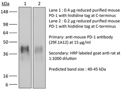
in vivo blocking of PD-1/PD-L signaling in vitro PD-1 neutralization Immunohistochemistry (frozen) Immunofluorescence Western blot Flow cytometry in vitro Organoids/Organ-on-Chip |
| Formulation |
PBS, pH 7.0 Contains no stabilizers or preservatives |
| Endotoxin |
≤1EU/mg (≤0.001EU/μg) Determined by LAL assay |
| Purity |
≥95% Determined by SDS-PAGE |
| Sterility | 0.2 µm filtration |
| Production | Purified from cell culture supernatant in an animal-free facility |
| Purification | Protein G |
| RRID | AB_2687796 |
| Molecular Weight | 150 kDa |
| Storage | The antibody solution should be stored at the stock concentration at 4°C. Do not freeze. |
| Need a Custom Formulation? | See All Antibody Customization Options |
Application References
-
Gordon, S. R., et al (2017). "PD-1 expression by tumour-associated macrophages inhibits phagocytosis and tumour immunity" Nature 545(7655): 495-499.
PubMed
Programmed cell death protein 1 (PD-1) is an immune checkpoint receptor that is upregulated on activated T cells for the induction of immune tolerance. Tumour cells frequently overexpress the ligand for PD-1, programmed cell death ligand 1 (PD-L1), facilitating their escape from the immune system. Monoclonal antibodies that block the interaction between PD-1 and PD-L1, by binding to either the ligand or receptor, have shown notable clinical efficacy in patients with a variety of cancers, including melanoma, colorectal cancer, non-small-cell lung cancer and Hodgkin’s lymphoma. Although it is well established that PD-1-PD-L1 blockade activates T cells, little is known about the role that this pathway may have in tumour-associated macrophages (TAMs). Here we show that both mouse and human TAMs express PD-1. TAM PD-1 expression increases over time in mouse models of cancer and with increasing disease stage in primary human cancers. TAM PD-1 expression correlates negatively with phagocytic potency against tumour cells, and blockade of PD-1-PD-L1 in vivo increases macrophage phagocytosis, reduces tumour growth and lengthens the survival of mice in mouse models of cancer in a macrophage-dependent fashion. This suggests that PD-1-PD-L1 therapies may also function through a direct effect on macrophages, with substantial implications for the treatment of cancer with these agents.
-
Wang, W., et al (2018). "RIP1 Kinase Drives Macrophage-Mediated Adaptive Immune Tolerance in Pancreatic Cancer" Cancer Cell 34(5): 757-774 e757.
PubMed
Pancreatic ductal adenocarcinoma (PDA) is characterized by immune tolerance and immunotherapeutic resistance. We discovered upregulation of receptor-interacting serine/threonine protein kinase 1 (RIP1) in tumor-associated macrophages (TAMs) in PDA. To study its role in oncogenic progression, we developed a selective small-molecule RIP1 inhibitor with high in vivo exposure. Targeting RIP1 reprogrammed TAMs toward an MHCII(hi)TNFalpha(+)IFNgamma(+) immunogenic phenotype in a STAT1-dependent manner. RIP1 inhibition in TAMs resulted in cytotoxic T cell activation and T helper cell differentiation toward a mixed Th1/Th17 phenotype, leading to tumor immunity in mice and in organotypic models of human PDA. Targeting RIP1 synergized with PD1-and inducible co-stimulator-based immunotherapies. Tumor-promoting effects of RIP1 were independent of its co-association with RIP3. Collectively, our work describes RIP1 as a checkpoint kinase governing tumor immunity.
-
Koyama, S., et al (2016). "STK11/LKB1 Deficiency Promotes Neutrophil Recruitment and Proinflammatory Cytokine Production to Suppress T-cell Activity in the Lung Tumor Microenvironment" Cancer Res 76(5): 999-1008.
PubMed
STK11/LKB1 is among the most commonly inactivated tumor suppressors in non-small cell lung cancer (NSCLC), especially in tumors harboring KRAS mutations. Many oncogenes promote immune escape, undermining the effectiveness of immunotherapies, but it is unclear whether the inactivation of tumor suppressor genes, such as STK11/LKB1, exerts similar effects. In this study, we investigated the consequences of STK11/LKB1 loss on the immune microenvironment in a mouse model of KRAS-driven NSCLC. Genetic ablation of STK11/LKB1 resulted in accumulation of neutrophils with T-cell-suppressive effects, along with a corresponding increase in the expression of T-cell exhaustion markers and tumor-promoting cytokines. The number of tumor-infiltrating lymphocytes was also reduced in LKB1-deficient mouse and human tumors. Furthermore, STK11/LKB1-inactivating mutations were associated with reduced expression of PD-1 ligand PD-L1 in mouse and patient tumors as well as in tumor-derived cell lines. Consistent with these results, PD-1-targeting antibodies were ineffective against Lkb1-deficient tumors. In contrast, treating Lkb1-deficient mice with an IL6-neutralizing antibody or a neutrophil-depleting antibody yielded therapeutic benefits associated with reduced neutrophil accumulation and proinflammatory cytokine expression. Our findings illustrate how tumor suppressor mutations can modulate the immune milieu of the tumor microenvironment, and they offer specific implications for addressing STK11/LKB1-mutated tumors with PD-1-targeting antibody therapies.
-
Koyama, S., et al (2016). "Adaptive resistance to therapeutic PD-1 blockade is associated with upregulation of alternative immune checkpoints" Nat Commun 7: 10501.
PubMed
Despite compelling antitumour activity of antibodies targeting the programmed death 1 (PD-1): programmed death ligand 1 (PD-L1) immune checkpoint in lung cancer, resistance to these therapies has increasingly been observed. In this study, to elucidate mechanisms of adaptive resistance, we analyse the tumour immune microenvironment in the context of anti-PD-1 therapy in two fully immunocompetent mouse models of lung adenocarcinoma. In tumours progressing following response to anti-PD-1 therapy, we observe upregulation of alternative immune checkpoints, notably T-cell immunoglobulin mucin-3 (TIM-3), in PD-1 antibody bound T cells and demonstrate a survival advantage with addition of a TIM-3 blocking antibody following failure of PD-1 blockade. Two patients who developed adaptive resistance to anti-PD-1 treatment also show a similar TIM-3 upregulation in blocking antibody-bound T cells at treatment failure. These data suggest that upregulation of TIM-3 and other immune checkpoints may be targetable biomarkers associated with adaptive resistance to PD-1 blockade.
Product Citations
-
Digital spatial profiling of α-PD-1 treated breast cancer bone metastases reveals region-specific signaling and enrichment of immune-suppressive markers.
In J Bone Oncol on 1 April 2026 by Grant, D. M., Joseph, G. J., et al.
PubMed
Bone is the most common and preferential site for breast cancer metastasis. Upon dissemination to the bone, breast cancer cells engraft into multiple niches, but it is unclear whether there are region-specific differences that may drive breast cancer progression in bone. We used a proteomic digital spatial profiling (DSP) approach to investigate which proliferation, cell death, and immune-related markers and pathways are enriched in immune and cancer cells residing 1) in the bone marrow or 2) along the endosteal surface, in an E0771, α-PD-1 treated pre-clinical model of breast cancer bone metastasis. We selected morphological markers to identify breast cancer cells and immune cells and applied a multiplexed set of probes targeting >70 proteins to characterize breast cancer and immune cell signaling in the marrow and endosteal regions using a DSP platform. We found multiple immune suppressive markers were enriched in the endosteum, including Foxp3, CD163, CD27, Pd-1, and Pd-l1, while proliferation markers were enriched in tumor cells in the marrow, including p38 Mapk, pan-Ras, Mek1, and phospho-Erk1/2. These findings shed light on the niche-specific proteins and pathways that are activated in breast cancer bone metastases and establish a user-friendly highly multiplexed approach for spatial proteomics in pre-clinical models of bone metastasis.
-
Curcumin enhances GSDME-mediated pyroptosis to potentiate PD-1/PD-L1 immune checkpoint blockade in colorectal cancer.
In Front Pharmacol on 18 February 2026 by Tan, D., Li, G., et al.
PubMed
Colorectal cancer (CRC) patients with a microsatellite-stable (MSS) status exhibit poor responsiveness to PD-1/PD-L1 blockade. Pyroptosis induction may resensitize MSS tumors to PD-1/PD-L1 blockade; however, the expression of GSDME, a key executor of pyroptosis, is often downregulated in CRC. Here, curcumin (CUR), a natural polyphenol, was identified as a potentiator of GSDME-dependent pyroptosis in CRC. We discovered that CUR upregulates GSDME expression by inhibiting the ubiquitin-proteasome system (UPS) in the MSS-type CT26 and HT29 cell lines and activating the caspase-3/GSDME signalling axis, resulting in increased pyroptosis. In CT26 tumors, CUR-enhanced pyroptosis reshaped tumor-infiltrating immune subsets and potentiated the efficacy of anti-PD-1 therapy. Notably, the synergistic antitumor activity of CUR combined with PD-1 blockade in CT26 tumors is strictly dependent on the caspase-3/GSDME axis, as the therapeutic benefit was abolished in GSDME-knockout tumors. These findings establish CUR as a safe and effective adjuvant for PD-1/PD-L1 blockade in MSS CRC, particularly in tumors with low GSDME expression.
-
Inhibiting OAS3 suppresses the development of clear cell renal cell carcinoma by reducing cell proliferation and altering the tumor immune microenvironment.
In Am J Transl Res on 12 February 2026 by Liao, X., Huang, P., et al.
PubMed
Immune checkpoint inhibitors have been approved for first-line treatment of metastatic clear cell renal cell carcinoma (ccRCC), but limited therapeutic effects have been reported in patients with advanced ccRCC. Investigating vital targets in specific immune interactions and their effect on the tumor microenvironment of ccRCC could provide novel strategies for overcoming the above limitations.
-
Multimodal spatial-omics reveal co-evolution of alveolar progenitors and proinflammatory niches in progression of lung precursor lesions.
In Cancer Cell on 9 February 2026 by Peng, F., Sinjab, A., et al.
PubMed
The co-evolution of different cell subsets in the progression of precursor lesions to lung adenocarcinoma (LUAD) is incompletely understood. We generated spatial transcriptomic maps of 56 human precursor lesions and LUADs from 25 patients and of an independent cohort of 36 lesions from 19 patients, analyzing a total of 486,519 spots and 5.4 million cells. We identify region-specific programs that distinguish precursors from LUADs. Spatially resolved clonal architectures reveal patient-specific heterogeneity in evolution of precursors to LUADs. We find epithelial alveolar progenitors expressing tumor-associated meta-programs and residing in niches enriched with proinflammatory subsets including IL1B high macrophages. Epithelial-proinflammatory niches are prevalent in precursor lesions but become less frequent in LUADs. These niches are conserved in mice and promote alveolar progenitor growth. Targeting inflammation alone or in combination with immune checkpoint blockade in precancerous phase reduces alveolar progenitors. Epithelial-inflammatory niches are stage-specific, shape early LUAD development and represent promising targets for interception.