
InVivoMAb anti-mouse PD-L1 (B7-H1)
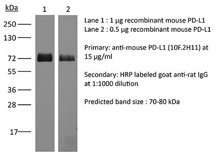
Product Description
Specifications
| Isotype | Rat IgG2b, κ |
|---|---|
| Recommended Isotype Control(s) | InVivoMAb rat IgG2b isotype control, anti-keyhole limpet hemocyanin |
| Recommended Dilution Buffer | InVivoPure pH 7.0 Dilution Buffer |
| Conjugation | This product is unconjugated. Conjugation is available via our Antibody Conjugation Services. |
| Immunogen | Murine PD-L1 expressing CHO cells |
| Reported Applications |
in vivo blocking of PD-L1/CD80 (B7-1) interactions in vitro blocking of PD-L1/CD80 (B7-1) interactions ELISA Flow cytometry |
| Formulation |
PBS, pH 7.0 Contains no stabilizers or preservatives |
| Endotoxin |
≤1EU/mg (≤0.001EU/μg) Determined by LAL assay |
| Purity |
≥95% Determined by SDS-PAGE |
| Sterility | 0.2 µm filtration |
| Purification | Protein G |
| RRID | AB_2927503 |
| Molecular Weight | 150 kDa |
| Storage | The antibody solution should be stored at the stock concentration at 4°C. Do not freeze. |
| Need a Custom Formulation? | See All Antibody Customization Options |
Application References
-
Mott, K. R., et al (2014). "Inclusion of CD80 in HSV targets the recombinant virus to PD-L1 on DCs and allows productive infection and robust immune responses" PLoS One 9(1): e87617.
PubMed
CD80 plays a critical role in stimulation of T cells and subsequent control of infection. To investigate the effect of CD80 on HSV-1 infection, we constructed a recombinant HSV-1 virus that expresses two copies of the CD80 gene in place of the latency associated transcript (LAT). This mutant virus (HSV-CD80) expressed high levels of CD80 and had similar virus replication kinetics as control viruses in rabbit skin cells. In contrast to parental virus, this CD80 expressing recombinant virus replicated efficiently in immature dendritic cells (DCs). Additionally, the susceptibility of immature DCs to HSV-CD80 infection was mediated by CD80 binding to PD-L1 on DCs. This interaction also contributed to a significant increase in T cell activation. Taken together, these results suggest that inclusion of CD80 as a vaccine adjuvant may promote increased vaccine efficacy by enhancing the immune response directly and also indirectly by targeting to DC.
-
Paterson, A. M., et al (2011). "The programmed death-1 ligand 1:B7-1 pathway restrains diabetogenic effector T cells in vivo" J Immunol 187(3): 1097-1105.
PubMed
Programmed death-1 ligand 1 (PD-L1) is a coinhibitory molecule that negatively regulates multiple tolerance checkpoints. In the NOD mouse model, PD-L1 regulates the development of diabetes. PD-L1 has two binding partners, programmed death-1 and B7-1, but the significance of the PD-L1:B7-1 interaction in regulating self-reactive T cell responses is not yet clear. To investigate this issue in NOD mice, we have compared the effects of two anti-PD-L1 Abs that have different blocking activities. Anti-PD-L1 mAb 10F.2H11 sterically and functionally blocks only PD-L1:B7-1 interactions, whereas anti-PD-L1 mAb 10F.9G2 blocks both PD-L1:B7-1 and PD-L1:programmed death-1 interactions. Both Abs had potent, yet distinct effects in accelerating diabetes in NOD mice: the single-blocker 10F.2H11 mAb was more effective at precipitating diabetes in older (13-wk-old) than in younger (6- to 7-wk-old) mice, whereas the dual-blocker 10F.9G2 mAb rapidly induced diabetes in NOD mice of both ages. Similarly, 10F.2H11 accelerated diabetes in recipients of T cells from diabetic, but not prediabetic mice, whereas 10F.9G2 was effective in both settings. Both anti-PD-L1 mAbs precipitated diabetes in adoptive transfer models of CD4(+) and CD8(+) T cell-driven diabetes. Taken together, these data demonstrate that the PD-L1:B7-1 pathway inhibits potentially pathogenic self-reactive effector CD4(+) and CD8(+) T cell responses in vivo, and suggest that the immunoinhibitory functions of this pathway may be particularly important during the later phases of diabetogenesis.
-
Yang, J., et al (2011). "The novel costimulatory programmed death ligand 1/B7.1 pathway is functional in inhibiting alloimmune responses in vivo" J Immunol 187(3): 1113-1119.
PubMed
The programmed death ligand 1 (PDL1)/programmed death 1 (PD1) costimulatory pathway plays an important role in the inhibition of alloimmune responses as well as in the induction and maintenance of peripheral tolerance. It has been demonstrated recently that PDL1 also can bind B7.1 to inhibit T cell responses in vitro. Using the bm12 into B6 heart transplant model, we investigated the functional significance of this interaction in alloimmune responses in vivo. PD1 blockade unlike PDL1 blockade failed to accelerate bm12 allograft rejection, suggesting a role for an additional binding partner for PDL1 other than PD1 in transplant rejection. PDL1 blockade was able to accelerate allograft rejection in B7.2-deficient recipients but not B7.1-deficient recipients, indicating that PDL1 interaction with B7.1 was important in inhibiting rejection. Administration of the novel 2H11 anti-PDL1 mAb, which only blocks the PDL1-B7.1 interaction, aggravated chronic injury of bm12 allografts in B6 recipients. Aggravated chronic injury was associated with an increased frequency of alloreactive IFN-gamma-, IL-4-, and IL-6-producing splenocytes and a decreased percentage of regulatory T cells in the recipients. Using an in vitro cell culture assay, blockade of the interaction of PDL1 on dendritic cells with B7.1 on T cells increased IFN-gamma production from alloreactive CD4(+) T cells, whereas blockade of dendritic cell B7.1 interaction with T cell PDL1 did not. These data indicate that PDL1 interaction with B7.1 plays an important role in the inhibition of alloimmune responses in vivo and suggests a dominant direction for PDL1 and B7.1 interaction.
Product Citations
-
Quantifying treatment response to a macrophage-targeted therapy in combination with immune checkpoint inhibitors after exposure to conventional chemotherapy.
In Front Immunol on 13 May 2025 by Bess, S. N., Smart, G. K., et al.
PubMed
Conventional chemotherapeutic agents, such as 5-fluorouracil (5-FU), can exert anti-tumor effects through immunogenic cell death (ICD) induction. Researchers have found hallmarks that quantify ICD (such as the translocation of HMGB1 and calreticulin). Although chemotherapeutic agents can induce ICD, they increase the expression of immune checkpoints, limiting their effectiveness. Studies have emphasized the importance of investigating the heterogeneous responses of cells co-localized in a solid tumor (macrophages, tumor cells, etc.) to ICD induction. However, these studies were performed in vivo, which limits the collection of information on cell-cell interactions due to model complexity.
-
Extracellular vesicle-packaged lncRNA from cancer-associated fibroblasts promotes immune evasion by downregulating HLA-A in pancreatic cancer.
In J Extracell Vesicles on 1 July 2024 by Yao, H., Huang, C., et al.
PubMed
Pancreatic ductal adenocarcinoma (PDAC) is characterised by immune evasion that contribute to poor prognosis. Cancer-associated fibroblasts (CAFs) play a pivotal role in orchestrating the PDAC tumour microenvironment. We investigated the role of CAF-derived extracellular vesicle (EV)-packaged long non-coding RNAs (lncRNAs) in immune evasion and explored gene therapy using engineered EVs loading small interfering RNAs (siRNAs) as a potential therapeutic strategy. Our findings highlight the significance of EV-packaged lncRNA RP11-161H23.5 from CAF in promoting PDAC immune evasion by downregulating HLA-A expression, a key component of antigen presentation. Mechanistically, RP11-161H23.5 forms a complex with CNOT4, a subunit of the mRNA deadenylase CCR4-NOT complex, enhancing the degradation of HLA-A mRNA by shortening its poly(A) tail. This immune evasion mechanism compromises the anti-tumour immune response. To combat this, we propose an innovative approach utilising engineered EVs as natural and biocompatible nanocarriers for siRNA-based gene therapy and this strategy holds promise for enhancing the effectiveness of immunotherapy in PDAC. Overall, our study sheds light on the critical role of CAF-derived EV-packaged lncRNA RP11-161H23.5/CNOT4/HLA-A axis in PDAC immune evasion and presents a novel avenue for therapeutic intervention.
-
Integrin αvβ6 mediates the immune escape through regulation of PD-L1 and serves as a novel marker for immunotherapy of colon carcinoma.
In Am J Cancer Res on 11 June 2024 by Yu, J., E, T., et al.
PubMed
The immune escape of colon cancer and its role in the response to immunotherapies such as PD-1/PD-L1 checkpoint inhibitors have long been of great interest. The positive outcomes of immunotherapy are limited by the immunosuppressive nature of the tumor microenvironment. Integrin αvβ6, which can regulate the progression of colon cancer, was recently reported to be involved in the immune suppression of colon cancer. In the present study, we explored the correlation between αvβ6 and PD-L1 expression by immunohistochemistry of colon cancer tissues. Then, the regulation of PD-L1 signaling by αvβ6 in colon cancer cells was demonstrated. We constructed an in vivo model and performed immunophenotyping experiments to analyze further the regulation of the immune response by αvβ6. The role of αvβ6 in the response to anti-PD-1 therapy in colon cancer was also verified. αvβ6-positive tissues exhibited increased PD-L1 expression. Inhibition of αvβ6 not only downregulated constitutive PD-L1 expression but also decreased IFN-γ-induced PD-L1 expression. In addition, αvβ6-induced PD-L1 expression was suppressed by the ERK inhibitor PD98059, and knockdown of the β6-ERK2 binding site had the equivalent effect. αvβ6 decreased CD8+ T cell infiltration and granzyme B expression in CD8+ T cells in colon cancer patients. Furthermore, mice engrafted with αvβ6-expressing colon cancer cells exhibited an unsatisfactory response to anti-PD-1 therapy, and anti-PD-1-induced increases in CD4+ and CD8+ T cell infiltration could be inhibited by αvβ6. These results indicate that αvβ6 mediates immune escape in colon cancer by upregulating PD-L1 through the ERK/MAPK pathway. Moreover, αvβ6 could serve as a marker for the efficacy of anti-PD-1 therapy in colon cancer.
-
Targeting the HSP47-collagen axis inhibits brain metastasis by reversing M2 microglial polarization and restoring anti-tumor immunity.
In Cell Rep Med on 21 May 2024 by Wang, L., Li, C., et al.
PubMed
Brain metastases (BrMs) are the leading cause of death in patients with solid cancers. BrMs exhibit a highly immunosuppressive milieu and poor response to immunotherapies; however, the underlying mechanism remains largely unclear. Here, we show that upregulation of HSP47 in tumor cells drives metastatic colonization and outgrowth in the brain by creating an immunosuppressive microenvironment. HSP47-mediated collagen deposition in the metastatic niche promotes microglial polarization to the M2 phenotype via the α2β1 integrin/nuclear factor κB pathway, which upregulates the anti-inflammatory cytokines and represses CD8+ T cell anti-tumor responses. Depletion of microglia reverses HSP47-induced inactivation of CD8+ T cells and abolishes BrM. Col003, an inhibitor disrupting HSP47-collagen association restores an anti-tumor immunity and enhances the efficacy of anti-PD-L1 immunotherapy in BrM-bearing mice. Our study supports that HSP47 is a critical determinant of M2 microglial polarization and immunosuppression and that blocking the HSP47-collagen axis represents a promising therapeutic strategy against brain metastatic tumors.