
InVivoMAb anti-mouse TNFα
in vitro TNFα neutralization
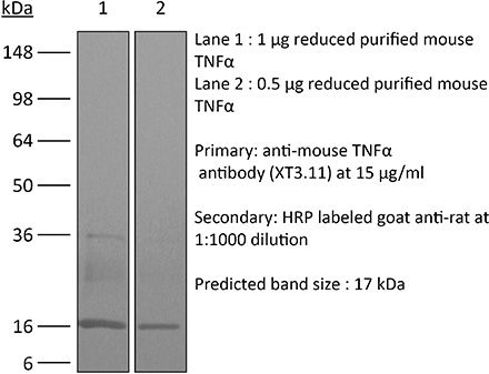
Western blot
Product Description
Specifications
| Isotype | Rat IgG1 |
|---|---|
| Recommended Isotype Control(s) | InVivoMAb rat IgG1 isotype control, anti-horseradish peroxidase |
| Recommended Dilution Buffer | InVivoPure pH 8.0 Dilution Buffer |
| Conjugation | This product is unconjugated. Conjugation is available via our Antibody Conjugation Services. |
| Immunogen | Recombinant mouse TNFα |
| Reported Applications |
in vivo TNFα neutralization in vitro TNFα neutralization Western blot |
| Formulation |
PBS, pH 8.0 Contains no stabilizers or preservatives |
| Endotoxin |
≤1EU/mg (≤0.001EU/μg) Determined by LAL assay |
| Purity |
≥95% Determined by SDS-PAGE |
| Sterility | 0.2 µm filtration |
| Production | Purified from cell culture supernatant in an animal-free facility |
| Purification | Protein G |
| RRID | AB_1107764 |
| Molecular Weight | 150 kDa |
| Storage | The antibody solution should be stored at the stock concentration at 4°C. Do not freeze. |
| Need a Custom Formulation? | See All Antibody Customization Options |
Application References
-
Deng, L., et al (2014). "Irradiation and anti-PD-L1 treatment synergistically promote antitumor immunity in mice" J Clin Invest 124(2): 687-695.
PubMed
High-dose ionizing irradiation (IR) results in direct tumor cell death and augments tumor-specific immunity, which enhances tumor control both locally and distantly. Unfortunately, local relapses often occur following IR treatment, indicating that IR-induced responses are inadequate to maintain antitumor immunity. Therapeutic blockade of the T cell negative regulator programmed death-ligand 1 (PD-L1, also called B7-H1) can enhance T cell effector function when PD-L1 is expressed in chronically inflamed tissues and tumors. Here, we demonstrate that PD-L1 was upregulated in the tumor microenvironment after IR. Administration of anti-PD-L1 enhanced the efficacy of IR through a cytotoxic T cell-dependent mechanism. Concomitant with IR-mediated tumor regression, we observed that IR and anti-PD-L1 synergistically reduced the local accumulation of tumor-infiltrating myeloid-derived suppressor cells (MDSCs), which suppress T cells and alter the tumor immune microenvironment. Furthermore, activation of cytotoxic T cells with combination therapy mediated the reduction of MDSCs in tumors through the cytotoxic actions of TNF. Our data provide evidence for a close interaction between IR, T cells, and the PD-L1/PD-1 axis and establish a basis for the rational design of combination therapy with immune modulators and radiotherapy.
-
Shaabani, N., et al (2018). "The probacterial effect of type I interferon signaling requires its own negative regulator USP18" Sci Immunol 3(27).
PubMed
Type I interferon (IFN-I) signaling paradoxically impairs host immune responses during many primary and secondary bacterial infections. Lack of IFN-I receptor reduces bacterial replication and/or bacterial persistence during infection with several bacteria. However, the mechanisms that mediate the adverse IFN-I effect are incompletely understood. Here, we show that Usp18, an interferon-stimulated gene that negatively regulates IFN-I signaling, is primarily responsible for the deleterious effect of IFN-I signaling during infection of mice with Listeria monocytogenes or Staphylococcus aureus Mechanistically, USP18 promoted bacterial replication by inhibiting antibacterial tumor necrosis factor-alpha (TNF-alpha) signaling. Deleting IFNAR1 or USP18 in CD11c-Cre(+) cells similarly reduced bacterial titers in multiple organs and enhanced survival. Our results demonstrate that inhibiting USP18 function can promote control of primary and secondary bacterial infection by enhancing the antibacterial effect of TNF-alpha, which correlates with induction of reactive oxygen species (ROS). These findings suggest that USP18 could be targeted therapeutically in patients to ameliorate disease caused by serious bacterial infections.
-
Baeyens, A., et al (2015). "Effector T cells boost regulatory T cell expansion by IL-2, TNF, OX40, and plasmacytoid dendritic cells depending on the immune context" J Immunol 194(3): 999-1010.
PubMed
CD4(+)CD25(+)Foxp3(+) regulatory T (Treg) cells play a major role in peripheral tolerance. Multiple environmental factors and cell types affect their biology. Among them, activated effector CD4(+) T cells can boost Treg cell expansion through TNF or IL-2. In this study, we further characterized this effector T (Teff) cell-dependent Treg cell boost in vivo in mice. This phenomenon was observed when both Treg and Teff cells were activated by their cognate Ag, with the latter being the same or different. Also, when Treg cells highly proliferated on their own, there was no additional Treg cell boost by Teff cells. In a condition of low inflammation, the Teff cell-mediated Treg cell boost involved TNF, OX40L, and plasmacytoid dendritic cells, whereas in a condition of high inflammation, it involved TNF and IL-2. Thus, this feedback mechanism in which Treg cells are highly activated by their Teff cell counterparts depends on the immune context for its effectiveness and mechanism. This Teff cell-dependent Treg cell boost may be crucial to limit inflammatory and autoimmune responses.
-
Christensen, A. D., et al (2015). "Depletion of regulatory T cells in a hapten-induced inflammation model results in prolonged and increased inflammation driven by T cells" Clin Exp Immunol 179(3): 485-499.
PubMed
Regulatory T cells (Tregs ) are known to play an immunosuppressive role in the response of contact hypersensitivity (CHS), but neither the dynamics of Tregs during the CHS response nor the exaggerated inflammatory response after depletion of Tregs has been characterized in detail. In this study we show that the number of Tregs in the challenged tissue peak at the same time as the ear-swelling reaches its maximum on day 1 after challenge, whereas the number of Tregs in the draining lymph nodes peaks at day 2. As expected, depletion of Tregs by injection of a monoclonal antibody to CD25 prior to sensitization led to a prolonged and sustained inflammatory response which was dependent upon CD8 T cells, and co-stimulatory blockade with cytotoxic T lymphocyte antigen-4-immunoglobulin (CTLA-4-Ig) suppressed the exaggerated inflammation. In contrast, blockade of the interleukin (IL)-10-receptor (IL-10R) did not further increase the exaggerated inflammatory response in the Treg -depleted mice. In the absence of Tregs , the response changed from a mainly acute reaction with heavy infiltration of neutrophils to a sustained response with more chronic characteristics (fewer neutrophils and dominated by macrophages). Furthermore, depletion of Tregs enhanced the release of cytokines and chemokines locally in the inflamed ear and augmented serum levels of the systemic inflammatory mediators serum amyloid (SAP) and haptoglobin early in the response.
Product Citations
-
TNF-mediated hilar interneuron loss and aberrant granule cell migration are associated with chronic cognitive deficits following TBI.
In Brain Behav Immun on 1 March 2026 by Harris, E. A., Budianto, S., et al.
PubMed
Chronic morbidities, including cognitive impairment, are a common consequence of traumatic brain injury (TBI), with millions currently living with permanent TBI-related disabilities. Recent work has indicated that altered cellular architecture in the dentate gyrus (DG) may play a significant role in the development of chronic cognitive impairment and excitotoxicity. However, current understanding of the temporal progression of these pathological changes in the context of neuroinflammation and chronic cognitive outcomes is limited. This study characterized temporospatial changes in the hilar region of the DG, showing that the population of reelin- and somatostatin-expressing inhibitory interneurons was significantly reduced as early as 7 days post-injury (dpi), and that aberrant migration of excitatory granule cells occurs gradually in the weeks to months following injury. These findings coincided with upregulation of monocyte/macrophage-associated inflammatory mediators, including MIP-1β, MIG, MCP-1, and TNF-α at 7 days dpi, with differential cytokine regulation persisting 120 dpi. Injury was associated with the development of chronic spatial memory impairment and reduced risk-assessment behavior, with a transient reduction in spontaneous anxiety. TNFR1 and TNFR2 were differentially expressed in inhibitory neurons, further implicating TNF-signaling as a driver of hilar neuron loss. Furthermore, systemic administration of anti-TNF-α monoclonal antibody induced significant neuroprotection, attenuated pro-inflammatory mediators, and hilar interneuron loss. These findings suggest that TNF-TNFR signaling plays a crucial role in driving hilar interneuron loss and aberrant granule cell migration, which, in turn, may contribute to the development of excitotoxicity and chronic cognitive deficits.
-
TNF-mediated hilar interneuron loss and aberrant granule cell migration are associated with chronic cognitive deficits following TBI.
In Brain Behav Immun on 1 March 2026 by Harris, E. A., Budianto, S., et al.
PubMed
Chronic morbidities, including cognitive impairment, are a common consequence of traumatic brain injury (TBI), with millions currently living with permanent TBI-related disabilities. Recent work has indicated that altered cellular architecture in the dentate gyrus (DG) may play a significant role in the development of chronic cognitive impairment and excitotoxicity. However, current understanding of the temporal progression of these pathological changes in the context of neuroinflammation and chronic cognitive outcomes is limited. This study characterized temporospatial changes in the hilar region of the DG, showing that the population of reelin- and somatostatin-expressing inhibitory interneurons was significantly reduced as early as 7 days post-injury (dpi), and that aberrant migration of excitatory granule cells occurs gradually in the weeks to months following injury. These findings coincided with upregulation of monocyte/macrophage-associated inflammatory mediators, including MIP-1β, MIG, MCP-1, and TNF-α at 7 days dpi, with differential cytokine regulation persisting 120 dpi. Injury was associated with the development of chronic spatial memory impairment and reduced risk-assessment behavior, with a transient reduction in spontaneous anxiety. TNFR1 and TNFR2 were differentially expressed in inhibitory neurons, further implicating TNF-signaling as a driver of hilar neuron loss. Furthermore, systemic administration of anti-TNF-α monoclonal antibody induced significant neuroprotection, attenuated pro-inflammatory mediators, and hilar interneuron loss. These findings suggest that TNF-TNFR signaling plays a crucial role in driving hilar interneuron loss and aberrant granule cell migration, which, in turn, may contribute to the development of excitotoxicity and chronic cognitive deficits.
-
Acute peritonitis-induced adipose CD127+ ILC1s express PD-L1 and ameliorate inflammation in mice.
In Nat Commun on 5 February 2026 by Nagata, R., Akama, Y., et al.
PubMed
Peritonitis is an inflammation of the peritoneum primarily caused by gut perforation and consequent bacterial leakage, a known cause of sepsis. Although adipose tissue is recognized as an immunologically active organ, the involvement of adipose tissue innate lymphoid cells (ILC) in regulating peritonitis remains poorly understood. Here, we employ a cecal ligation and puncture mouse model and demonstrate that circulating CD127- group 1 ILC (ILC1) migrate into the mesenteric adipose tissue (MAT) during the inflammatory period of peritonitis. CD127- ILC1s undergo phenotypic changes to become CD127+ ILC1s, resulting in an increased number of CD127+ ILC1s in the MAT. We also show that this population of CD127+ ILC1s expresses PD-L1, exhibits low IFN-γ production, and potentially acts as a negative regulator of TNF production by γδ T cells, thereby controlling acute peritonitis. Our findings suggest that MAT-CD127+ ILC1s play an important regulatory role in acute peritonitis and may represent a potential therapeutic target for sepsis.
-
CARD14 signaling in intestinal epithelial cells induces intestinal inflammation and intestinal transit delay.
In EMBO Mol Med on 1 December 2025 by Aidarova, A., Carels, M., et al.
PubMed
CARD14 is an intracellular NF-κB signaling mediator in the skin, and rare CARD14 variants have been associated with psoriasis and atopic dermatitis. CARD14 is also expressed in intestinal epithelial cells (IEC). However, its function in the intestine remains unknown. We demonstrate here that transgenic mice expressing the psoriasis-associated gain-of-function human CARD14(E138A) mutant specifically in IEC show mild intestinal inflammation, without epithelial damage. Moreover, CARD14(E138A)IEC mice show a drastic reduction in intestinal motility, often associated with rectal prolapse. Enteric neuronal survival and functionality are unaffected in CARD14(E138A)IEC mice. Transcriptome analysis of IEC from CARD14(E138A)IEC mice reveals decreased expression of antimicrobial peptides by Paneth cells, accompanied by microbial dysbiosis and increased susceptibility to enteric bacterial infection. Our findings suggest that gain-of-function CARD14 mutations may not only predispose patients to psoriasis but also mild intestinal inflammation, reduced intestinal motility, and increased sensitivity to intestinal infection. CARD14(E138A)IEC mice are also a valuable tool for further investigation of IEC-intrinsic molecular processes involved in intestinal inflammation and motility disorders.