
InVivoMAb anti-mouse/rat IL-1β
in vitro IL-1β neutralization
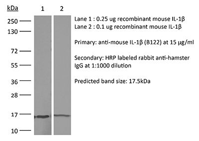
ELISA
Product Description
Specifications
| Isotype | Armenian hamster IgG |
|---|---|
| Recommended Isotype Control(s) | InVivoMAb polyclonal Armenian hamster IgG |
| Recommended Dilution Buffer | InVivoPure pH 7.0 Dilution Buffer |
| Conjugation | This product is unconjugated. Conjugation is available via our Antibody Conjugation Services. |
| Immunogen | Recombinant mouse IL-1β |
| Reported Applications |
in vivo IL-1β neutralization in vitro IL-1β neutralization ELISA |
| Formulation |
PBS, pH 7.0 Contains no stabilizers or preservatives |
| Endotoxin |
≤1EU/mg (≤0.001EU/μg) Determined by LAL assay |
| Purity |
≥95% Determined by SDS-PAGE |
| Sterility | 0.2 µm filtration |
| Production | Purified from cell culture supernatant in an animal-free facility |
| Purification | Protein G |
| RRID | AB_2687727 |
| Molecular Weight | 150 kDa |
| Storage | The antibody solution should be stored at the stock concentration at 4°C. Do not freeze. |
| Need a Custom Formulation? | See All Antibody Customization Options |
Application References
-
Coffelt, S. B., et al (2015). "IL-17-producing gammadelta T cells and neutrophils conspire to promote breast cancer metastasis" Nature 522(7556): 345-348.
PubMed
Metastatic disease remains the primary cause of death for patients with breast cancer. The different steps of the metastatic cascade rely on reciprocal interactions between cancer cells and their microenvironment. Within this local microenvironment and in distant organs, immune cells and their mediators are known to facilitate metastasis formation. However, the precise contribution of tumour-induced systemic inflammation to metastasis and the mechanisms regulating systemic inflammation are poorly understood. Here we show that tumours maximize their chance of metastasizing by evoking a systemic inflammatory cascade in mouse models of spontaneous breast cancer metastasis. We mechanistically demonstrate that interleukin (IL)-1beta elicits IL-17 expression from gamma delta (gammadelta) T cells, resulting in systemic, granulocyte colony-stimulating factor (G-CSF)-dependent expansion and polarization of neutrophils in mice bearing mammary tumours. Tumour-induced neutrophils acquire the ability to suppress cytotoxic T lymphocytes carrying the CD8 antigen, which limit the establishment of metastases. Neutralization of IL-17 or G-CSF and absence of gammadelta T cells prevents neutrophil accumulation and downregulates the T-cell-suppressive phenotype of neutrophils. Moreover, the absence of gammadelta T cells or neutrophils profoundly reduces pulmonary and lymph node metastases without influencing primary tumour progression. Our data indicate that targeting this novel cancer-cell-initiated domino effect within the immune system–the gammadelta T cell/IL-17/neutrophil axis–represents a new strategy to inhibit metastatic disease.
-
Khmaladze, I., et al (2014). "Mannan induces ROS-regulated, IL-17A-dependent psoriasis arthritis-like disease in mice" Proc Natl Acad Sci U S A 111(35): E3669-3678.
PubMed
Psoriasis (Ps) and psoriasis arthritis (PsA) are poorly understood common diseases, induced by unknown environmental factors, affecting skin and articular joints. A single i.p. exposure to mannan from Saccharomyces cerevisiae induced an acute inflammation in inbred mouse strains resembling human Ps and PsA-like disease, whereas multiple injections induced a relapsing disease. Exacerbation of disease severity was observed in mice deficient for generation of reactive oxygen species (ROS). Interestingly, restoration of ROS production, specifically in macrophages, ameliorated both skin and joint disease. Neutralization of IL-17A, mainly produced by gammadelta T cells, completely blocked disease symptoms. Furthermore, mice depleted of granulocytes were resistant to disease development. In contrast, certain acute inflammatory mediators (C5, Fcgamma receptor III, mast cells, and histamine) and adaptive immune players (alphabeta T and B cells) were redundant in disease induction. Hence, we propose that mannan-induced activation of macrophages leads to TNF-alpha secretion and stimulation of local gammadelta T cells secreting IL-17A. The combined action of activated macrophages and IL-17A produced in situ drives neutrophil infiltration in the epidermis and dermis of the skin, leading to disease manifestations. Thus, our finding suggests a new mechanism triggered by exposure to exogenous microbial components, such as mannan, that can induce and exacerbate Ps and PsA.
-
Chen, K. W., et al (2014). "The neutrophil NLRC4 inflammasome selectively promotes IL-1beta maturation without pyroptosis during acute Salmonella challenge" Cell Rep 8(2): 570-582.
PubMed
The macrophage NLRC4 inflammasome drives potent innate immune responses against Salmonella by eliciting caspase-1-dependent proinflammatory cytokine production (e.g., interleukin-1beta [IL-1beta]) and pyroptotic cell death. However, the potential contribution of other cell types to inflammasome-mediated host defense against Salmonella was unclear. Here, we demonstrate that neutrophils, typically viewed as cellular targets of IL-1beta, themselves activate the NLRC4 inflammasome during acute Salmonella infection and are a major cell compartment for IL-1beta production during acute peritoneal challenge in vivo. Importantly, unlike macrophages, neutrophils do not undergo pyroptosis upon NLRC4 inflammasome activation. The resistance of neutrophils to pyroptotic death is unique among inflammasome-signaling cells so far described and allows neutrophils to sustain IL-1beta production at a site of infection without compromising the crucial inflammasome-independent antimicrobial effector functions that would be lost if neutrophils rapidly lysed upon caspase-1 activation. Inflammasome pathway modification in neutrophils thus maximizes host proinflammatory and antimicrobial responses during pathogen challenge.
-
Gopinath, S., et al (2014). "Role of disease-associated tolerance in infectious superspreaders" Proc Natl Acad Sci U S A 111(44): 15780-15785.
PubMed
Natural populations show striking heterogeneity in their ability to transmit disease. For example, a minority of infected individuals known as superspreaders carries out the majority of pathogen transmission events. In a mouse model of Salmonella infection, a subset of infected hosts becomes superspreaders, shedding high levels of bacteria (>10(8) cfu per g of feces) but remain asymptomatic with a dampened systemic immune state. Here we show that superspreader hosts remain asymptomatic when they are treated with oral antibiotics. In contrast, nonsuperspreader Salmonella-infected hosts that are treated with oral antibiotics rapidly shed superspreader levels of the pathogen but display signs of morbidity. This morbidity is linked to an increase in inflammatory myeloid cells in the spleen followed by increased production of acute-phase proteins and proinflammatory cytokines. The degree of colonic inflammation is similar in antibiotic-treated superspreader and nonsuperspreader hosts, indicating that the superspreader hosts are tolerant of antibiotic-mediated perturbations in the intestinal tract. Importantly, neutralization of acute-phase proinflammatory cytokines in antibiotic-induced superspreaders suppresses the expansion of inflammatory myeloid cells and reduces morbidity. We describe a unique disease-associated tolerance to oral antibiotics in superspreaders that facilitates continued transmission of the pathogen.
Product Citations
-
GSDME/IL-18 pyroptotic axis prevents myosteatosis by expanding tissue-resident macrophages to promote muscle regeneration.
In J Clin Invest on 17 February 2026 by Cao, Q., Liu, J., et al.
PubMed
Metabolic-inflammatory crosstalk orchestrates muscle repair. Although pyroptosis typically aggravates sterile injury, we demonstrated that GSDME-dependent pyroptotic signaling associated with recruited myeloid cells paradoxically supported regeneration. GSDME expression was induced in post-surgical human muscle injury and murine damage models. Gsdme deficiency delayed functional recovery and exacerbated injury-induced myosteatosis, a pathological form of intramuscular ectopic fat deposition. Time-series and single-cell RNA-sequencing analyses revealed that GSDME loss shifted the transcriptional program from oxidative metabolism toward lipid storage and adipogenesis. Lipidomics confirmed aberrant accumulation of triacylglycerols and sphingolipids in Gsdme-deficient muscle. Single-cell profiling further identified divergent fibro-adipogenic progenitors (FAPs) states skewed toward adipogenesis, accompanied by impaired expansion of restorative Lyve1⁺Cd163⁺Txnip⁺ tissue-resident macrophages (TRMs)-validated by multiplex flow cytometry. Blocking CCR2-dependent monocyte recruitment produced regenerative defects comparable to those caused by Gsdme deficiency. Myeloid-specific Gsdme reintroduction rescued TRM expansion and function, curbed FAP adipogenic reprogramming, whereas FAP-specific expression proved ineffective. Mechanistically, IL-18 downstream of GSDME-dependent signaling engaged KLF4/JUN signaling in TRMs, sustaining their reparative and lipid-clearing capacity. This GSDME-IL-18-TRMs axis was compromised in aged muscle, yet exogenous IL-18 reversed myosteatosis and accelerated regeneration. Together, these findings suggest that GSDME-dependent pyroptotic signaling can act as a metabolic checkpoint that sustains TRM-driven lipid homeostasis to support muscle regeneration.
-
Deficiency of lysosomal TMEM175 in myeloid macrophages exerts anti-tumor immunity via inflammasome and cross-presentation pathway.
In Nat Commun on 14 February 2026 by Zhang, Z., Li, X., et al.
PubMed
Discovering more targets is of great importance for developing alternative interventions for tumor therapy. The roles of transmembrane protein 175 (TMEM175) in neurodegeneration diseases have been reported, however its functions in tumor immune surveillance are not known. We show that TMEM175 conditional knockout in macrophages inhibits the tumor growth and metastasis through promoting the anti-tumor immunity in the tumor microenvironment (TME), including elevated M1-like polarization, reduced M2-like polarization, and facilitated recruitment and activation of T cells and nature killer cells (NKs). The anti-tumor immunity is abrogated by caspase-1 inhibitor VX-765, anti-IL-1β, and anti-IL-18. Tmem175-/- bone marrow-derived macrophages (BMDMs) show enhanced tumor antigen cross-presentation that is further strengthened by IL-1β and IL-18. NLRP3 is robustly elicited in Tmem175-/- BMDMs by the tumor cell debris through lysosomal permeabilization and cathepsin B leakage. Finally, Tmem175-/- mice are more responsive to anti-PD-1. Our works implies TMEM175 to be a potential target for immunotherapy.
-
Tet2 deficiency-induced expansion of monocyte-derived macrophages promotes liver fibrosis.
In J Exp Med on 2 February 2026 by Feng, J., Wu, B., et al.
PubMed
Clonal hematopoiesis driven by Tet2 deficiency in myeloid cells (TetΔMye) is prevalent in elderly individuals; however, the role of Tet2ΔMye in liver fibrosis pathogenesis remains elusive. In this study, we demonstrated that Tet2-deficient monocyte-derived macrophages (MDMs) promoted cellular expansion and elevated C-C motif chemokine ligand 2/8 (Ccl2/8) secretion by stabilizing their mRNAs through 5hmC-mediated alterations in RNA-protein interactions. These chemokines engaged with the upregulated C-C motif chemokine receptor (Ccr2/3) on Tet2-/- monocytes, forming a positive feedback loop that amplified pro-inflammatory MDMs (pMDMs) accumulation in liver. Tet2-/- pMDMs activated hepatic stellate cells through IL-6, driving extracellular matrix deposition and fibrotic progression. Pharmacological inhibition of Ccl2/Ccl8 with Bindarit attenuated MDMs accumulation and liver fibrosis, whereas combined therapy with Bindarit and IL-6 neutralization synergistically suppressed liver fibrosis in Tet2ΔMye mice and aged chimeric models recapitulating Tet2ΔMye-related myeloid hematopoiesis. These findings present the mechanism that Tet2ΔMye aggravates liver fibrosis and highlight MDMs depletion plus IL-6 neutralization as a promising therapy for liver fibrosis in patients with Tet2ΔMye-related myeloid hematopoiesis.
-
The Ly6ghigh Neutrophil Subset Dictates Breast Cancer Lung Metastasis via CD8+ T Cell Death.
In Cancer Commun (Lond) on 2 February 2026 by Wang, R., Liu, X., et al.
PubMed
Background: Lung metastasis is a leading cause of breast cancer (BC)-related mortality, driven by the immunosuppressive traits of the metastatic tumor microenvironment. However, the mechanisms underlying cell-cell crosstalk in shaping immune evasion within the metastatic niche remain poorly defined. Neutrophil extracellular traps (NETs) and their associated proteins, such as cathelicidin, have emerged as key mediators of metastatic regulation in cancer. Here, we aimed to decipher the interaction between a neutrophil subset characterized by high expression of lymphocyte antigen 6 complex locus g (Ly6ghigh) and cluster of differentiation 8-positive T lymphocytes (CD8+ T cells), mediated via cathelicidin embedded in NETs, as well as their synergistic mechanism and cooperative role in promoting lung metastasis of BC. Methods: We characterized neutrophil heterogeneity and functional dynamics by performing single-cell RNA sequencing and flow cytometry on lung tissues derived from murine models of BC lung metastasis. We utilized cathelicidin-related antimicrobial peptide (Cramp) knockout mice to dissect the role of cathelicidin in NETs. The spatial colocalization of apoptotic CD8+ T cells and NETs was analyzed using multiplex immunofluorescence, and the molecular interactions were probed by protein binding assays. Results: Neutrophils in the lung metastatic niche were classified into 2 subsets based on the Ly6g expression: Ly6ghigh and Ly6glow neutrophils. Ly6glow neutrophils, which were recruited in the macrometastatic stage, exhibited myeloid-derived suppressor cell-like characteristics. Notably, Ly6ghigh neutrophils induced CD8+ T cell apoptosis through NET formation, with apoptotic CD8+ T cells spatially clustered within NET-rich areas. Mechanistically, NET-derived cathelicidin (Cramp in mice) directly bound to mitochondrial adenine nucleotide translocator 1 (Ant1) in CD8+ T cells, triggering conformational changes and complex formation with voltage-dependent anion channel 1 (Vdac1). These events resulted in the opening of the mitochondrial permeability transition pore and loss of mitochondrial membrane potential. Conclusions: Our study demonstrates that Ly6ghigh neutrophils play a critical role in immunosuppression and immune evasion through NET-induced apoptosis of CD8+ T cells. These findings underscore the importance of NETs and cathelicidin in BC lung metastasis, suggesting their potential as therapeutic targets in restoring antitumor immunity and in preventing metastatic progression.