
InVivoMAb anti-mouse IL-4
in vitro IL-4 neutralization
in vivo IL-4 receptor stimulation (as a complex with IL-4)
Flow cytometry
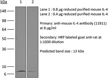
Western blot
Product Description
Specifications
| Isotype | Rat IgG1, κ |
|---|---|
| Recommended Isotype Control(s) | InVivoMAb rat IgG1 isotype control, anti-horseradish peroxidase |
| Recommended Dilution Buffer | InVivoPure pH 7.0 Dilution Buffer |
| Conjugation | This product is unconjugated. Conjugation is available via our Antibody Conjugation Services. |
| Immunogen | Partially purified native mouse IL-4 |
| Reported Applications |
in vivo IL-4 neutralization in vitro IL-4 neutralization in vivo IL-4 receptor stimulation (as a complex with IL-4) Flow cytometry Western blot |
| Formulation |
PBS, pH 7.0 Contains no stabilizers or preservatives |
| Endotoxin |
≤1EU/mg (≤0.001EU/μg) Determined by LAL assay |
| Purity |
≥95% Determined by SDS-PAGE |
| Sterility | 0.2 µm filtration |
| Production | Purified from cell culture supernatant in an animal-free facility |
| Purification | Protein G |
| RRID | AB_1107707 |
| Molecular Weight | 150 kDa |
| Storage | The antibody solution should be stored at the stock concentration at 4°C. Do not freeze. |
| Need a Custom Formulation? | See All Antibody Customization Options |
Application References
-
Burton, B. R., et al (2014). "Sequential transcriptional changes dictate safe and effective antigen-specific immunotherapy" Nat Commun 5: 4741.
PubMed
Antigen-specific immunotherapy combats autoimmunity or allergy by reinstating immunological tolerance to target antigens without compromising immune function. Optimization of dosing strategy is critical for effective modulation of pathogenic CD4(+) T-cell activity. Here we report that dose escalation is imperative for safe, subcutaneous delivery of the high self-antigen doses required for effective tolerance induction and elicits anergic, interleukin (IL)-10-secreting regulatory CD4(+) T cells. Analysis of the CD4(+) T-cell transcriptome, at consecutive stages of escalating dose immunotherapy, reveals progressive suppression of transcripts positively regulating inflammatory effector function and repression of cell cycle pathways. We identify transcription factors, c-Maf and NFIL3, and negative co-stimulatory molecules, LAG-3, TIGIT, PD-1 and TIM-3, which characterize this regulatory CD4(+) T-cell population and whose expression correlates with the immunoregulatory cytokine IL-10. These results provide a rationale for dose escalation in T-cell-directed immunotherapy and reveal novel immunological and transcriptional signatures as surrogate markers of successful immunotherapy.
-
Tang, W., et al (2014). "The oncoprotein and transcriptional regulator Bcl-3 governs plasticity and pathogenicity of autoimmune T cells" Immunity 41(4): 555-566.
PubMed
Bcl-3 is an atypical member of the IkappaB family that modulates transcription in the nucleus via association with p50 (NF-kappaB1) or p52 (NF-kappaB2) homodimers. Despite evidence attesting to the overall physiologic importance of Bcl-3, little is known about its cell-specific functions or mechanisms. Here we demonstrate a T-cell-intrinsic function of Bcl-3 in autoimmunity. Bcl-3-deficient T cells failed to induce disease in T cell transfer-induced colitis and experimental autoimmune encephalomyelitis. The protection against disease correlated with a decrease in Th1 cells that produced the cytokines IFN-gamma and GM-CSF and an increase in Th17 cells. Although differentiation into Th1 cells was not impaired in the absence of Bcl-3, differentiated Th1 cells converted to less-pathogenic Th17-like cells, in part via mechanisms involving expression of the RORgammat transcription factor. Thus, Bcl-3 constrained Th1 cell plasticity and promoted pathogenicity by blocking conversion to Th17-like cells, revealing a unique type of regulation that shapes adaptive immunity.
-
Gu, A. D., et al (2015). "A critical role for transcription factor Smad4 in T cell function that is independent of transforming growth factor beta receptor signaling" Immunity 42(1): 68-79.
PubMed
Transforming growth factor-beta (TGF-beta) suppresses T cell function to maintain self-tolerance and to promote tumor immune evasion. Yet how Smad4, a transcription factor component of TGF-beta signaling, regulates T cell function remains unclear. Here we have demonstrated an essential role for Smad4 in promoting T cell function during autoimmunity and anti-tumor immunity. Smad4 deletion rescued the lethal autoimmunity resulting from transforming growth factor-beta receptor (TGF-betaR) deletion and compromised T-cell-mediated tumor rejection. Although Smad4 was dispensable for T cell generation, homeostasis, and effector function, it was essential for T cell proliferation after activation in vitro and in vivo. The transcription factor Myc was identified to mediate Smad4-controlled T cell proliferation. This study thus reveals a requirement of Smad4 for T-cell-mediated autoimmunity and tumor rejection, which is beyond the current paradigm. It highlights a TGF-betaR-independent role for Smad4 in promoting T cell function, autoimmunity, and anti-tumor immunity.
-
Clever, D., et al (2016). "Oxygen Sensing by T Cells Establishes an Immunologically Tolerant Metastatic Niche" Cell 166(5): 1117-1131 e1114.
PubMed
Cancer cells must evade immune responses at distant sites to establish metastases. The lung is a frequent site for metastasis. We hypothesized that lung-specific immunoregulatory mechanisms create an immunologically permissive environment for tumor colonization. We found that T-cell-intrinsic expression of the oxygen-sensing prolyl-hydroxylase (PHD) proteins is required to maintain local tolerance against innocuous antigens in the lung but powerfully licenses colonization by circulating tumor cells. PHD proteins limit pulmonary type helper (Th)-1 responses, promote CD4(+)-regulatory T (Treg) cell induction, and restrain CD8(+) T cell effector function. Tumor colonization is accompanied by PHD-protein-dependent induction of pulmonary Treg cells and suppression of IFN-gamma-dependent tumor clearance. T-cell-intrinsic deletion or pharmacological inhibition of PHD proteins limits tumor colonization of the lung and improves the efficacy of adoptive cell transfer immunotherapy. Collectively, PHD proteins function in T cells to coordinate distinct immunoregulatory programs within the lung that are permissive to cancer metastasis.
Product Citations
-
A fungi-derived cyclic peptide enhances Th9-mediated antitumor immunity by targeting ZAP70 and SREBP1.
In J Clin Invest on 2 February 2026 by Zhao, W., Zhou, Y., et al.
PubMed
Adoptive cell therapy (ACT) relies on durable and functional T cells to mediate tumor clearance. Th9 cells are a metabolically fit CD4+ T cell subset with strong persistence but limited cytotoxicity. Here, we identified endomelipeptide A (EpA), a cyclic peptide isolated from Ganoderma lucidum-associated endophytic fungi, as a potent enhancer of Th9 cell differentiation. EpA promoted a cytotoxic Th9 phenotype with enhanced mitochondrial function and metabolic fitness. Mechanistically, EpA dually targeted ZAP70 and SREBP1, coupling T cell receptor signaling activation with lipid metabolism suppression. EpA-treated Th9 cells mediated robust, CD8+ T cell-dependent tumor control and enhanced the efficacy of human Th9 CAR T cell therapy in vivo. These findings establish EpA as a distinct cyclic peptide that reprograms Th9 cells and provides a potential approach to boost ACT efficacy.
-
CCL20-CCR6 signaling alters the metabolic reprogramming to promote the pathogenic Th17 cell differentiation.
In iScience on 16 January 2026 by Meitei, H. T., Kulkarni, N., et al.
PubMed
CCR6 is a G-protein-coupled receptor that binds to its ligand CCL20. Th17 and Foxp3+CD4+ T cells express CCR6, enabling their migration to inflamed tissues with unique metabolic environments. The factors that regulate their metabolic adaptation and functional roles in these tissues remain unclear. Using inflammatory bowel disease patient samples and experimental colitis models in mice, we demonstrated that the intrinsic signaling of CCL20-CCR6 in CD4+ T cells promotes the differentiation of inflammatory Th1-like Th17 cells (T-bet+RORγt+) during colitis. This signaling induces the rapamycin-sensitive phosphorylation of PI3K, Akt, mTORC1, and STAT3 in a CCR6-dependent manner. RNA-seq and proteomics analysis revealed that the addition of CCL20 during Th17 differentiation affects several metabolic pathways, including energy metabolism. CCL20 significantly increased glycolysis and inhibited oxidative phosphorylation, thereby driving the differentiation of pathogenic Th17 cells. Our findings suggest that alterations in CCR6-induced changes in Th17 metabolism offer an interesting therapeutic target for gut inflammation and autoimmunity.
-
The ionotropic AMPA receptor contributes to autoimmunity via altered regulatory T cell differentiation.
In iScience on 16 January 2026 by Mitchell-Flack, M., Higgins, M., et al.
PubMed
The AMPA receptor (AMPAR) is an ionotropic glutamate receptor that is essential for neuronal communication, yet its role in the immune system remains poorly understood. Here, using a CD4Cre selective deletion mouse model, we provide the first functional characterization of AMPAR deficient T cells. We demonstrate that AMPAR deletion in T cells significantly protects against severe paralysis in an experimental autoimmune encephalomyelitis (EAE) model, and this protection is associated with increased regulatory T cell (Treg) presence within the spinal cord. In vitro studies reveal that the deletion of the AMPAR intrinsically promotes Treg generation. Mechanistically, AMPAR deletion increases IL2 signaling and activates the mTORC1 pathway, supporting Treg development and function. These novel findings suggest that a function of the AMPAR in CD4 T cells is to limit immune suppression by restricting Treg differentiation. Targeting AMPARs on T cells could offer a novel therapeutic approach for the treatment of autoimmune disease.